| Catalog_no | AP0093 |
| Product_name | eIF2α (phospho Ser51) Polyclonal Antibody |
| Category | 抗原抗体 |
| Applications | IF,WB,IHC-p,ELISA |
| Reactivity | Human,Mouse,Rat |
| Size | 100μg/50μg/20μg |
| Price | 2200.00/1200.00/560.00 |
| Gene_name | EIF2S1 |
| Protein_name | Eukaryotic translation initiation factor 2 subunit 1 |
| Human swiss prot no | 1965 |
| Human swiss prot no | P05198 |
| Mouse gene id | 13665 |
| Mouse swiss prot no | Q6ZWX6 |
| Rat gene id | 54318 |
| Rat swiss prot no | P68101 |
| immunogen | The antiserum was produced against synthesized peptide derived from human eIF2 alpha around the phosphorylation site of Ser51. AA range:21-70 |
| Specificity | Phospho-eIF2α (S51) Polyclonal Antibody detects endogenous levels of eIF2α protein only when phosphorylated at S51. |
| Formulation | Liquid in PBS containing 50% glycerol, 0.5% BSA and 0.02% sodium azide. |
| Source | Rabbit |
| Dilution | IF: 1:50-200 Western Blot: 1/500 - 1/2000. Immunohistochemistry: 1/100 - 1/300. ELISA: 1/10000. Not yet tested in other applications. |
| Purification | The antibody was affinity-purified from rabbit antiserum by affinity-chromatography using epitope-specific immunogen. |
| Concentration | 1 mg/ml |
| Storage_stability | -20°C/1 year |
| Msds | MSDS_Antibody.pdf |
| othername | EIF2S1; EIF2A; Eukaryotic translation initiation factor 2 subunit 1; Eukaryotic translation initiation factor 2 subunit alpha; eIF-2-alpha; eIF-2A; eIF-2alpha |
| Observed_band | 38KD |
| Instructions | |
| Share |
if-rat-heart119.jpg) |
Immunofluorescence analysis of rat-heart tissue. 1,eIF2α (phospho Ser51) Polyclonal Antibody(red) was diluted at 1:200(4°C,overnight). 2, Cy3 labled Secondary antibody was diluted at 1:300(room temperature, 50min).3, Picture B: DAPI(blue) 10min. Picture A:Target. Picture B: DAPI. Picture C: merge of A+B |
if-rat-heart120.jpg) |
Immunofluorescence analysis of rat-heart tissue. 1,eIF2α (phospho Ser51) Polyclonal Antibody(red) was diluted at 1:200(4°C,overnight). 2, Cy3 labled Secondary antibody was diluted at 1:300(room temperature, 50min).3, Picture B: DAPI(blue) 10min. Picture A:Target. Picture B: DAPI. Picture C: merge of A+B |
 |
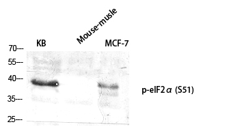
Western Blot analysis of various cells using Phospho-eIF2α (S51) Polyclonal Antibody diluted at 1:2000 |
.jpg) |
Western Blot analysis of 293 cells using Phospho-eIF2α (S51) Polyclonal Antibody diluted at 1:2000 |


Copyright © 2021 苏州博特龙免疫技术有限公司 版权所有 ,苏ICP备2021013855号-1
苏公网安备32050702011981号 